Friday, February 18, 2011

POWERED SURGICAL INSTRUMENTS


SURGICAL ULTRASONIC EQUIPMENT
Instrument using high energy source for surgery.
It works through ease of dissection, and aspiration of soft tissue

- Ultrasonic techniques are well established in neurosurgery for rapid and efficient removal of intracranial and spinal cord tumors.
- This instrument is also used in resection, and dissection applications of:

(a) Liver,
(b) Kidney, and
(c) Spleen.

- Their application is also gaining popularity in pancreatic and urologic surgery.
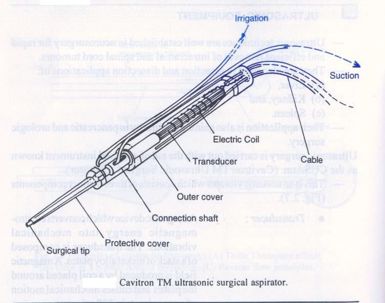
Ultrasonic surgery is carried out with the aid of a special instrument known as the CUSAtm (Cavitron TM Ultrasonic Surgical Aspirator).
- This is an acoustic vibrator which consists of three distinct components.

• Transducer: A device, that converts electromagnetic energy into mechanical vibrations. The transducer is composed of a stack of nickel alloy plates. A magnetic field is produced by a coil placed around the plates and causes mechanical motion of approximately 300 microns.
• Connecting body: Mechanically conveys the motions of the transducer to the surgical tip. It also amplifies the vibration motion of the transducer.
• Surgical tip: Completes the amplifications of the motion and also contacts the tissue. For this reason the tip is relatively long compared to its diameter and this provides adequate motion amplification.

- The electric coil which is permanently fitted in the hand piece surrounds the transducer.
- This coil receives 23,000 cycles per second (hertz) alternating electric current from the console and activates the transducer.
- The hand piece is connected to the console by a cable which includes the tubing for circulating fluid between the cooling water canister in the console and the hand piece.
- Since the electric coil has a current flowing through it heat is generated and absorbed by the water circulating within the hand piece.
- This keeps the hand piece at a comfortable temperature for the surgeon.
Any questions be sent to drmmkapur@gmail.com
All earlier posts are stored in archives for access and review.
Vistors that follow the site may post contributions to the site.
Click on image tosee details.

Thursday, February 10, 2011

HIGH INTENSITY LIGHT


LASERS

Lasers are an equipment that use high energy to destroy, or seal tissue

- A laser is an instrument equipped to produce a fine parallel beam of high intensity light, which can be focused on a very small spot.
- The development of surgical lasers, during the last decade, has made a significant contribution to microsurgery.
- Used with an operating microscope or fiberscope a wide range of microsurgical procedures have now become possible, for example, suture less micro vascular anatomists.
- Excision of the posterior capsule of the lens through the transparent cornea.
- Coagulation of bleeding peptic ulcers using a flexible gastro scope.
- In laser surgery, the light beam heats the target tissue and causes:

• Thermal(heat) tissue destruction, first with local edema and then by denaturing of proteins.
• Contraction of tissue due to alteration of fibrous tissue protein and finally boiling of cell water and vaporization.
• Generally, low energy exposure results in coagulation of blood vessel.
• Higher energy exposure produces a precise incision by vaporization of the tissue at the focal point.
Any questions be sent to drmmkapur@gmail.com
All earlier posts are stored in archives for your access and review.
Visitors that follow the site may post contributions to the site.
Click on image to see details.

Thursday, February 3, 2011

COOL POWERED INSTRUMENT


CRYOSURGICAL SYSTEMS

Cryosurgical is the application of extreme cold to tissues and is a useful technique for the destruction of tissues.
It is characterized by minimal bleeding, or pain, in the post-operative period.
Cryosurgical procedures are well established in many specialties including:

• General surgery,
• Dermatology,
• Neurology, and
• Urology.


The first practical equipment for the control and maintenance of extreme low temperature in surgery was produced in 1962.
The apparatus was not very sophisticated and used liquid nitrogen to achieve low temperature in the region of – 196°C.
This very low temperature was subsequently found to exceed requirements.

A more simple apparatus which used nitrous oxide or carbon dioxide to achieve temperature in the region of – 70°C.
The design principles established, are still used in modern cryosurgical equipment which employs high-pressure, non-siphon cylinders of N2O, or CO2.

- Cryosurgical systems consist of a flexible tubing, connecting the gas flow control unit, to a cryoprobe or working tip, this unit comprises of two concentric tubes.
- The inner tube delivers nitrous oxide or carbon dioxide at pressures between 4,000 and 6,000 kpa to a narrow orifice in the end.
- The sudden expansion of this gas, through the Joule-Thompson orifice, produces a rapid drop in temperature of the probe surface, forming an ice ball.
- The expanded gas is then returned at atmospheric pressure along the outer tube.
- Some cooling along the shaft of the probe occurs, which can be a disadvantage, for example in neurosurgery.
- To overcome this, manufactures designed a probe which employs a reversed gas flow.
- This design enables the incoming gas, to be carried to the probe tip, via the outer of the two concentric tubes; it is allowed to expand through an annular orifice, being finally returned through the central tube.
- The incoming gas acts as an insulating barrier and the cooling is confined to the probe tip.
Any questions be sent to drmmkapur@gmail.com
All earlier posts are stored in archives for your access and review
Visitors that follow the site may post contributions to the site.
Click on image to see details.

Friday, January 28, 2011

POWERED INSTRUMENTS


ELECTRIC INSTRUMENTS


SURGICAL CAUTERY

In surgery we require to burn tissue with heat, cautery serves this limited purpose well.
With battery power it can be used at home or in the office

- An electric cautery consists of a platinum wire loop or point which becomes red hot when a current is passed through it by an electrode.
- This heated cautery point is then applied to the tissue area to cause tissue death through coagulation.
- The current source may be a low-voltage battery or a transformer connected to the mains; the transformer is preferable, as the output is more constant than that of a battery.
- The cautery must not be used any hotter than at red heat, as too high a current will cause rapid burning out of the cautery wire.
- The cautery Point are of various sizes and shapes and are mounted on a heat-resistant handle to which are connected to wires.
- The cautery must be connected to the cautery terminals.
- After connecting the wires the transformer may then be switched on.
Ay questions be sent to drmmkapur@gmail.com
All earlier posts are stored in archives for access and review.
Visitors that follow the site may post contributions to the site.
Click on image to see details

Friday, January 21, 2011

POWERED INSTRUMENTS


Electric Instruments

Mono-polar diathermy(cont)


The purpose of the indifferent large surface electrode in mono-polar diathermy, is to provide a surface of sufficient area to avoid any heat effects at this site.

- This is because the indifferent electrode is in contact with hundreds and thousands of more cells than the live electrode which results in a lower density of current in each of these cells.
- If the electrode is applied correctly as described below; little heat is generated in this area.
- The commonest type of indifferent electrode is a flexible metal plate. The thin flexible metal plate electrode can either be placed under the patient’s body (sacrum) or carefully bandaged round the thigh to ensure even contact with the skin.
- It is necessary to use electrode jelly, if the patient is hairy, shaving beforehand is advisable as hair acts as an electrical resistance.
- If the patient’s skin is dry as in our dry climate, moistening with water or saline can be used with advantage before applying the plate. The plate can be wrapped in a moist sheet.
- Replace a plate electrode which becomes uneven, otherwise areas of irregular contact with the skin result, which could allow points of high current density and risk of burns.
- The electrode is connected to the high frequency electrosurgical unit indifferent or earth terminal by a heavy insulated wire.
- Risk of accidents is possible.
Bipolar diathermy is a safer alternative.


With a bipolar system the surgeon’s instrument, usually in the form of a forceps, Combines both electrodes so that current flows down both limbs of the forceps
Only the tissue gripped between the tips of the forceps receives the current.
Eliminating the need for an indifferent electrode.
Any questions be sent to drmmkapur@gmail.com
All earlier posts are stored in archives for access and review
Visitors that follow the site may post contributions to the site
Click on image to see details

Friday, January 14, 2011

POWER INSTRUMENTS IN SURGERY


ELECTRIC INSTRUMENTS


Diathermy
Diathermy machines generate high frequency electric current.
This energy can be used by the surgeon to cut skin and deeper tissues.
It can also be used to coagulate tissue to limit bleeding.
These effects are the result of heat generated in the tissues


Surgical diathermy is frequently used in surgery to limit bleeding..
- When this current is passed through the patient’s body between two electrodes, the effect is to produce a concentration of current at the smaller electrode in the hand of the surgeon.
- As the surgeon applies his live (small) electrode to the tissues, the current passes through the touched tissue cells and owing to the tissue’s electrical resistance, heat is generated at this contact point.
- The effect is localized because with mono-polar system the current from the live (small) electrode spreads out in the patient’s body and travels to the indifferent electrode which is a large electrode placed in contact with the patient’s body (thigh or sacrum).
- A high density of current occurs only immediately beneath the live electrode because further away (except under fault conditions) the current density is too small to have any heating effect.
- Fault conditions are produced by the contact of patients body with the metallic table
Any questins be sent to drmmkapur@gmail.com
All earlier posts are stored in archives for access and review.
Visitors that follow the site may post contributions to the site.
Click in image to see details

Friday, January 7, 2011

THE FINAL SCENE


We show here some views thru the endoscopes
The upper GI endoscopy shows views of stomach mucosa
The lower GI shows colon and removal of polyps


WHAT ARE THE DIFFERENT GASTROINTESTINAL FIBRESCOPES (INCLUDING GASTRSCOPES, DUODENOSCOPES AND CLONOSCOPES)?

Based on the angle of view are three types of instruments:-

1. Forward or oblique (for oblique) viewing for examination and
biopsy(a)
This is suitable for examining the oesophagus, most of the
stomach and the upper part of the duodenum
2. Side or lateral viewing for examination, biopsy and operative
procedures in the duodenum(b)
This is needed when full examination of oesphagus, stomach,
duodenum and biliary system is required. This includes the
fundus of the stomach and the duodenal bulb
3. Forward viewing with two channels for complex operative
procedures.
This is generally larger in diameter than the previous two and
has suitable dimension channels incorporated for operating
instruments. A wide range of procedures can be undertaken(c)

All three types of instruments can be obtained from several
manufacturers
Any questions be sent to drmmkapur@gmail.com
All earlier posts are stored in archives for access and review.
Visitors that follow may post contributions and comments
Click on image to see details